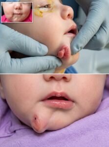
In 2020, doctors documented an extraordinary medical case involving a baby girl born with an extremely rare facial abnormality: a tiny, second mouth-like structure developing beside her normal mouth.
The condition was so unusual that it immediately attracted medical attention. Yet behind the remarkable images and complex diagnosis was something much simpler—a newborn baby who needed careful treatment, protection, and the chance to grow normally.
Her story began before she was even born.
Doctors Noticed Something Unusual Before Birth
During pregnancy, prenatal imaging revealed an unexpected mass near the right side of the baby’s developing lower jaw.
At that stage, doctors could see that something unusual was developing, but they couldn’t immediately determine what it was. Several possibilities had to be considered, including a cyst, a benign growth, or another congenital abnormality.
Prenatal imaging can reveal many developmental differences before birth, but it doesn’t always provide a complete picture of what doctors will find after delivery.
So the medical team prepared for the possibility that the newborn might require additional examinations once she arrived.
When the baby was eventually born full-term, however, doctors discovered something far more unusual than they had initially expected.
A Tiny Structure That Looked Like Another Mouth
The newborn was otherwise doing well.
She could breathe normally.
She could feed.
Her airway was stable.
But beside her lower jaw was a small structure that looked remarkably similar to a miniature mouth.
The unusual formation included tissue resembling a small lip and a tongue-like structure.
Even more remarkable was the movement doctors observed.
When the baby’s normal tongue moved, the tiny additional tongue-like structure also appeared to move.
For the medical team, this was an extraordinary finding because it suggested that the additional structure wasn’t simply an isolated piece of tissue. It appeared to contain several components associated with normal oral development.
But doctors knew that what they could see externally was only part of the story.
They needed to understand what was happening underneath the skin.
Advanced Scans Revealed More
Doctors used detailed imaging, including CT and MRI scans, to examine the baby’s facial anatomy.
The scans revealed that the unusual structure contained partially developed bone.
But there was another astonishing discovery.
Inside the additional structure were several developing teeth.
That meant the condition involved much more than an unusual external growth. Multiple types of tissue associated with the normal development of a mouth and jaw had developed in an additional location.
Doctors identified the condition as an extremely rare form of craniofacial duplication.
Craniofacial duplication refers to developmental abnormalities in which certain facial structures become duplicated to varying degrees.
Importantly, this baby did not have two complete faces.
Her primary facial structures had developed normally. Instead, the duplication was localized to an area beside her lower jaw, involving portions of oral tissue and jaw-related structures.
Why Did This Happen?
One of the most fascinating questions surrounding rare congenital conditions is why they happen in the first place.
The development of a human face is an incredibly complicated biological process.
Long before a baby is born, cells are moving, dividing, and forming specialized tissues. Bones begin developing. Muscles take shape. Nerves connect with tissues. The jaw forms. The structures that eventually become teeth begin developing.
All of these processes have to happen in the correct locations and at the correct stages.
Usually, this remarkable sequence unfolds with extraordinary precision.
But occasionally, development takes an unexpected path.
In cases involving extremely rare facial duplication, scientists and doctors may not be able to identify one simple cause. The underlying mechanisms can be complex and may involve unusual developmental processes occurring very early in pregnancy.
For this little girl, however, the immediate concern wasn’t discovering exactly why it happened.
The first priority was making sure she was safe.
Could She Breathe and Eat Normally?
Doctors needed to answer several important questions.
Was the additional structure connected to her normal mouth?
Did it communicate with her airway?
Could it interfere with feeding?
Could it affect the development of her normal jaw?
And would leaving it untreated create problems as she grew?
Fortunately, the baby’s condition was relatively stable.
The additional structure did not significantly interfere with her main oral cavity or airway. She was able to breathe normally and feed successfully.
That was extremely important.
Because she was stable, doctors didn’t have to rush into an emergency operation immediately after birth.
Instead, they had something incredibly valuable: time.
The medical team could study the anatomy carefully and plan the safest possible procedure.
Waiting Until She Was Six Months Old
After evaluating the baby’s condition, doctors decided to delay surgery until she was approximately six months old.
This allowed specialists to better understand the relationship between the duplicated tissues and her normal jaw and facial structures.
Operating on a newborn’s face requires extraordinary precision. The surgeons had to consider developing bones, muscles, nerves, blood vessels, salivary structures, and the baby’s future facial growth.
The goal wasn’t simply to remove the unusual structure.
They also needed to preserve the normal anatomy and reconstruct the affected area as carefully as possible.
For the family, those six months must have been filled with uncertainty and questions.
But the baby’s ability to breathe and feed normally provided an encouraging foundation while doctors prepared for treatment.
The Operation
At approximately six months old, the baby underwent surgery.
The medical team carefully removed the additional oral structure and reconstructed the affected portion of the lower jaw and surrounding soft tissue.
The operation required detailed knowledge of the unusual anatomy because the extra structure wasn’t made of just one type of tissue.
After removal, examination of the tissue revealed just how remarkable the duplication had been.
The structure contained muscle, salivary-gland tissue, dental tissue, and multiple developing teeth.
What had looked from the outside like a tiny second mouth was actually a complex collection of partially developed facial structures.
For the surgeons and medical specialists involved, it provided an extraordinary example of how different tissues can develop together in an unexpected location.
A Hopeful Recovery
The most important news came after surgery.
The little girl recovered well.
She continued to feed successfully, and follow-up examinations showed an encouraging overall outcome.
Doctors did report some continuing weakness affecting movement of part of her lower lip, but the overall result was positive.
The surgery had successfully addressed the unusual duplicated structure while allowing the baby to continue growing and developing.
For her family, the procedure represented much more than a successful medical operation.
It offered a chance to move forward without the unusual structure becoming a major obstacle to her development.
More Than a Rare Medical Case
It would be easy to look at this story simply as an unusual medical curiosity.
A baby born with what appeared to be a second mouth is certainly something most people have never seen.
But there is another side to the story.
Behind every rare medical case is a real person.
This wasn’t simply “the baby with a second mouth.”
She was someone’s daughter.
She was a newborn who needed to be fed, comforted, protected, and loved.
Her doctors weren’t treating her as a curiosity. They were trying to understand her unique anatomy so they could give her the safest and healthiest future possible.
That distinction matters.
Medical photographs can be shocking or fascinating, but they represent real human beings and real families.
Her story is therefore not only about an extraordinary congenital condition. It is also about compassion, medical expertise, and the importance of treating every patient with dignity.
Why Her Case Matters to Medicine
Extremely rare cases can provide valuable information to the medical community.
When doctors encounter an unusual congenital condition, documenting the patient’s imaging, anatomy, treatment, and recovery can help other specialists recognize similar cases in the future.
A doctor may go years without seeing a particular abnormality.
Then, years later, another baby may be born with a similar developmental difference.
Published medical case reports can give physicians valuable information about what to look for, how to investigate the condition, and what treatment options may be considered.
In that sense, this little girl’s case contributed to medical knowledge far beyond her own treatment.
Her unusual anatomy offered doctors a rare glimpse into the complicated processes that shape the human face before birth.
A Remarkable Beginning
Before she was born, doctors saw an unexplained mass.
After delivery, they discovered a tiny structure resembling another mouth.
Advanced imaging revealed bone and developing teeth.
Specialists studied the unusual anatomy and carefully planned a treatment.
Six months later, surgeons removed the duplicated structure and reconstructed the affected area.
And most importantly, the little girl recovered and continued growing.
Her journey demonstrates how far modern medicine has come.
Conditions that once might have been impossible to understand can now be examined using advanced imaging, studied in extraordinary detail, and treated through specialized surgery.
But perhaps the biggest lesson isn’t about technology.
It’s about perspective.
Being born with an extremely rare condition doesn’t make someone less deserving of a normal childhood.
This little girl needed what every child needs: care, patience, protection, and the opportunity to grow.
Her condition made her medically extraordinary, but it didn’t define who she was.
A Story of Science, Compassion, and Hope
The human body is capable of extraordinary things.
Most of the time, development follows a predictable path. But occasionally, nature takes an unexpected turn, creating conditions that challenge even experienced medical professionals.
For this little girl, that unexpected path resulted in an extraordinarily rare craniofacial condition.
Yet her story didn’t end with the diagnosis.
Doctors investigated.
They studied.
They waited for the safest moment.
They performed a complex operation.
And she recovered.
What began as uncertainty before birth ultimately became a story of understanding and treatment.
Her case reminds us that behind every extraordinary medical photograph is a human story—and that curiosity should always be accompanied by compassion.
At only six months old, this little girl underwent a procedure that gave her a new beginning.
She wasn’t defined by the tiny second mouth she was born with.
She was simply a child who received the care she needed and the opportunity to continue growing.
And sometimes, that is the most powerful part of any medical story: not how unusual the beginning was, but how much hope can come after it.
Full story in the comment below 👇❤️
Shared for medical education and awareness. Rare congenital conditions can vary significantly, and diagnosis, treatment, and outcomes depend on each patient’s individual anatomy and medical needs.